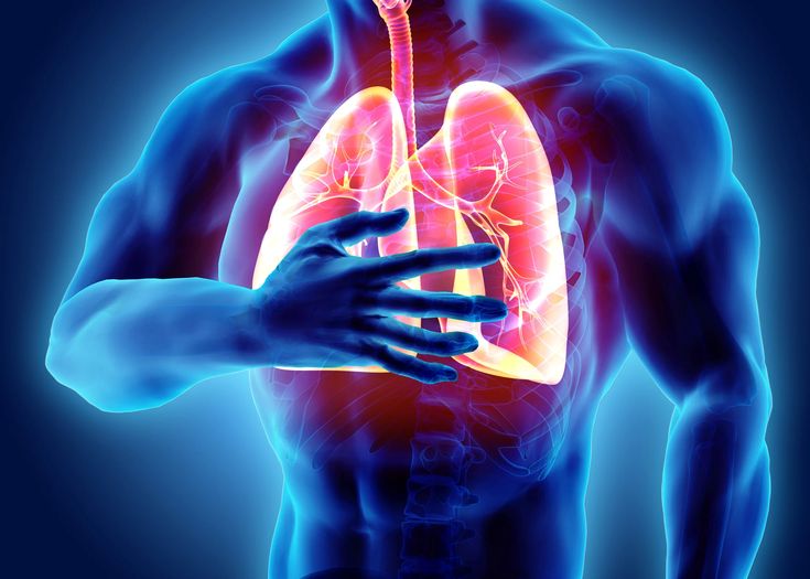
Introduction to Pulmonary Fibrosis
Pulmonary fibrosis is a chronic, progressive lung disease characterized by thickening and scarring (fibrosis) of lung tissue, making it difficult to breathe. Over time, the stiffened lung tissue loses its ability to transfer oxygen into the bloodstream, leading to respiratory failure.
Key Facts
Type: Interstitial lung disease (ILD)
Progression: Irreversible scarring worsens over time
Survival Rate: 3-5 years average after diagnosis (varies by type)
Prevalence: Affects ~100,000 people in the U.S., with 50,000 new cases yearly
Causes & Risk Factors
Known Causes
✔ Long-term exposure to toxins (asbestos, coal dust, silica)
✔ Chronic autoimmune diseases (RA, lupus, sarcoidosis)
✔ Radiation therapy (for lung/breast cancer)
✔ Certain medications (chemotherapy, heart drugs like amiodarone)
Risk Factors
Age: Most patients are 50+ years old
Smoking: Increases risk of IPF
Genetics: Family history raises susceptibility
GERD: Chronic acid reflux may worsen lung damage
Types of Pulmonary Fibrosis
1. Idiopathic Pulmonary Fibrosis (IPF)
Characteristics:
Most common and aggressive form
No identifiable cause (“idiopathic”)
Typically affects adults aged 50-70 years
Median survival: 3-5 years after diagnosis
Key Features:
Progressive scarring with honeycomb pattern on CT scans
More common in men and smokers
May have genetic predisposition in some cases
2. Hypersensitivity Pneumonitis (HP)
Characteristics:
Caused by repeated inhalation of allergens
Often occupational or hobby-related
Two forms: acute and chronic
Common Triggers:
Bird proteins (pigeon breeders’ disease)
Moldy hay (farmer’s lung)
Air conditioning/heating systems
Wood dust
3. Connective Tissue Disease-Associated PF
Associated Conditions:
Rheumatoid arthritis (most common)
Systemic sclerosis (scleroderma)
Sjögren’s syndrome
Lupus (SLE)
Polymyositis/dermatomyositis
Unique Aspects:
Often occurs alongside other symptoms (joint pain, skin changes)
May respond better to immunosuppressants than IPF
Prognosis varies by underlying condition
4. Occupational/Environmental PF
Major Causes:
Asbestosis: From asbestos exposure (construction, shipyards)
Silicosis: From silica dust (mining, stone cutting)
Coal Worker’s Pneumoconiosis: “Black lung” disease
Berylliosis: Aerospace/electronics industry exposure
Prevention Focus:
Proper workplace ventilation
Use of protective respirators
Regular health screenings for at-risk workers
5. Drug-Induced PF
Common Culprits:
Chemotherapy drugs (bleomycin, methotrexate)
Heart medications (amiodarone)
Certain antibiotics (nitrofurantoin)
Biological agents (rituximab)
Management:
Early recognition is critical
Often reversible if caught early
Requires medication review by specialists
6. Radiation-Induced PF
Occurs After:
Radiation therapy for lung, breast, or esophageal cancer
Typically develops 6-24 months post-treatment
Risk Factors:
Higher radiation doses
Combined chemotherapy
Pre-existing lung disease
7. Genetic Forms of PF
Inherited Conditions:
Familial pulmonary fibrosis
Hermansky-Pudlak syndrome
Telomere-related disorders
Genetic Testing:
Recommended for patients with family history
May influence treatment choices
Important for family counseling
Symptoms of Pulmonary Fibrosis
Early Signs
Dry, hacking cough (no mucus)
Shortness of breath (initially only during exercise)
Fatigue and weakness
Advanced Symptoms
Rapid, shallow breathing
Clubbing (enlarged fingertips/nails)
Unexplained weight loss
Chest discomfort (tightness or pain)
When to See a Doctor
⚠ Seek immediate care if:
Oxygen levels drop below 90%
Extreme breathlessness at rest
Blue lips/fingernails (cyanosis)
Diagnosis & Testing
1. Imaging Tests
High-Resolution CT Scan: Shows honeycomb-like scarring
Chest X-ray: Rules out other conditions
2. Lung Function Tests
Spirometry: Measures airflow restriction
Diffusion Capacity: Checks oxygen transfer ability
3. Biopsy (If Needed)
Surgical lung biopsy (for unclear cases)
Bronchoscopy (less invasive alternative)
4. Blood Tests
Check for autoimmune markers (ANA, RF)
Rule out infections
Treatment Options
1. Medications
Anti-fibrotic drugs (slow scarring):
Pirfenidone (Esbriet)
Nintedanib (Ofev)
Immunosuppressants (for autoimmune cases)
Antacids (if GERD contributes)
2. Oxygen Therapy
Portable oxygen tanks for daily use
Improves survival & quality of life
3. Pulmonary Rehabilitation
Breathing exercises
Physical therapy to maintain strength
4. Lung Transplant
Last resort for advanced cases
5-year survival rate: ~50-60%
Lifestyle & Home Remedies
Stop smoking (critical to slow progression)
Avoid pollutants (wear masks in dusty areas)
Vaccinations (flu, pneumonia, COVID-19)
Healthy diet (high-protein, antioxidant-rich foods)
Prognosis & Complications
Life Expectancy
Mild cases: 7-10 years
Severe IPF: 2-3 years without transplant
Common Complications
Respiratory failure (most common cause of death)
Pulmonary hypertension (high lung blood pressure)
Lung cancer (higher risk in long-term fibrosis)
Prevention Tips
Use protective gear if working with dust/chemicals
Manage autoimmune diseases aggressively
Regular lung check-ups if at high risk